Protocol for scoring TSR

TSR in epithelial carcinoma
The prognostic value of the TSR extends beyond colon cancer alone; it is observed in various other solid epithelial tumors. Like in colon cancer, in the other types of cancers, stroma-high tumors were associated with worse overall survival and disease-free survival. TSR is an independent prognostic factor in colon cancer, breast cancer, ovarian cancer, non-small cell lung cancer, nasopharyngeal cancer, esophageal cancer and hepatocellular cancer.
Moreover, similar to colon cancer, the TSR scoring method has a high interobserver agreement in a variety of studies on other epithelial cancer types, emphasizing the robustness of this method.
Tumor–stroma ratio
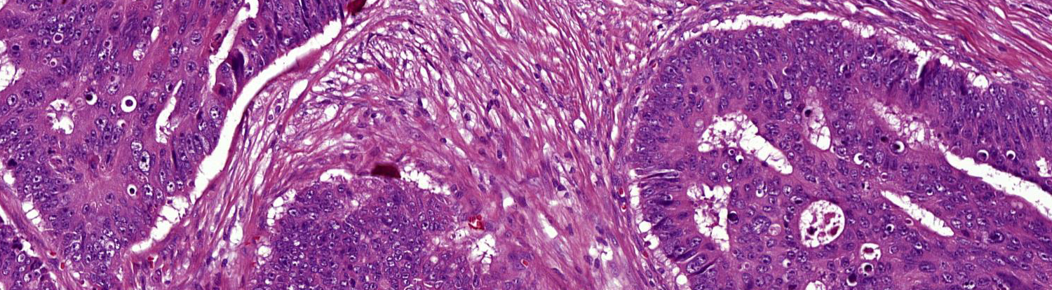
The tumor-stroma ratio (TSR) is based on the amount of stroma within the primary tumor. Several studies have shown the TSR to predict overall and disease-free survival in patients with colon carcinoma stage II and III. The TSR, combined with current routine pathohistology, can improve personalized treatment of these patients.
The TSR is a biomarker which can be easily determined in routine pathology. Scoring TSR only takes 1-2 minutes and is scored on the Haematoxylin & Eosin (H&E) stained sections of the tissue slide already used to determine the T-stadium of the tumor. There are no additional costs.
The parameter distinguishes tumors in two categories: Stroma-low tumors contain a stromal percentage of 50% or less (≤50%), whereas tumors containing more than 50% stroma, are called stroma-high. Patients with a stroma-high tumor have a worse overall and disease-free survival.


Exceptions
When determining the TSR, a number of aspects must be taken into account:
- Tumor epithelial cells should be present on all sides of the view;
- Score an area with as little necrosis, fatty or muscle tissue as possible, and preferably none at all. This is also the case for vessels and erythrocytes present in the tissue;
- In the case of mucinous tumors, an area containing mucus may be used. However, mucus should be excluded when determining the TSR.

E-learning
To participate in the UNITED study, the E-learning was necessary to correctly apprehend the scoring method. If a pathologist wanted to start the E-learning, they were sent the E-learning with instructions.
Once the E-learning was passed, a certificate was granted. After submission is completed and their center is participating, all certified pathologists received the rights and access to CastorEDC for inclusions.
References
See Research page.

Step 1: Select
Choose a section from the most invasive part of the tumor for microscopic analysis.

Step 2: Magnify
View with a 2.5x or 5x magnification and find the area with the most stroma visible.

Step 3: Zoom in
Zoom in and view that with a 10x magnification. Ensure that tumor cells are visible on all four sides of the area, and the least amount of other tissue as possible.

Step 4: Estimate percentage
Estimate a percentage for the amount of stroma, in increments of 10%.

Step 5: Determine and note
If there is more than one area with a potential highest amount of stroma, note the highest stromal percentage in the pathological report.
